
Mine Dosay-Akbulut
Department of Medical Biology and Genetics, Faculty of Veterinary, Afyon Kocatepe University, Afyon, Turkey
International Journal of Cancer Research
Year: 2006 | Volume: 2 | Issue: 2 | Page No.: 119-123


ABSTRACT
The world population and together problems especially the health problems are increase in every day. Cancer come a cross as one of the main important and uncured problem. A lot of countries including many developed countries such as USA release huge amounts of money towards cancer- related searches, aiming to find an effective treatment against to this problem. Every day brings new idea and solutions in finding the cure. In this study sharks, with a possible treatment link between shark and cancer mechanism, were presented as one of new source in the reality of unrecorded any cancer cases in the sharks up to know.
PDF Abstract XML References
How to cite this article
Mine Dosay-Akbulut, 2006. The Determination of the Specific Characteristics on the Immunosurveilance Against to Cancer Formation in Elasmobranchs. International Journal of Cancer Research, 2: 119-123.
DOI: 10.3923/ijcr.2006.119.123
URL: https://scialert.net/abstract/?doi=ijcr.2006.119.123
DOI: 10.3923/ijcr.2006.119.123
URL: https://scialert.net/abstract/?doi=ijcr.2006.119.123
INTRODUCTION
Cancer, uncontrolled growth of abnormal cell, which invade and destroy other tissues. Cancer develops in almost any organ or tissue of the body, but certain types of cancer are more lethal than others. Cancer is the leading cause of death in Canada and second only to heart disease in the United States. Each year, more than 1.2 million Americans and 125,000 Canadians are diagnosed with cancer, and about 622,000 North Americans-more than 1,700 people a day-die of the disease.
May be the worst cases of this century can be seen on cancer and aids. But people come across with cancer since beginning of the 19th century. Several different type of cancers, demands different treatments. Because up to now it hasn’t been found an effective treatments, even some of these research based on new sources. Up to know there hasn’t been recorded any cancer cases in sharks indicates a possible treatment link between shark and cancer mechanism.
There are several researches, indicates some shark-related materials, such as shark cartilage and its extracted materials, shark liver oil, shark’s high level of urea can play a role in the treatment against to the cancer and create a specific form in sharks body that somehow protect the sharks against to the cancer.
MATERIALS AND METHODS
DNA Sources, Amplification and Sequencing
The c-myc gene was chosen and sequenced for determining the nucleotide position of this oncogene, a possible reason formation of the cancer. This study was carried out in Quenn’s University of Belfast, School of Biology and Biochemistry Department, under the Michael Stanhope supervision between 1997-1998.
There are several reasons to being cancer. Cancer results either deragements of cell differentiation, or from mutations that disrupt the controls, regulating normal cell growth. The growth of normal cells is subject to many different types of control. Many oncogenes code for proteins that take part in various steps in the mechanism by which cells response to growth factors. Such oncogenes researches have been carried out for much better cancer treatments.
The c-myc or myc is a gene originally described in the avian MC29 myelocytomatosis virus, an oncovirus of the chicken. A homologous gene is located on the long arm of human chromosome 8. The translocation of coding sequence from its normal site in chromosome 8 to site on chromosome 14 or deletion of 5’ noncoding sequence of m-RNA can be the reason of the cancer in human. C-myc is also inducer of apoptosis. Both reductions of c-myc and inappropriate over-expression can be associated with cellular apoptosis and part of oncogenic process by enhancing cell proliferation and inhibition of cell differentiation (Panckham et al., 1994).
In the c-myc gene sequencing 4 species from different shark orders were used. They are Isurus oxyrinchus (shortfin mako) from order Lamniformes; Squatina californica (Pacific angel shark) from order Squatiniformes; Carcharhinus porosus (smalltail shark) from order Carcharhiniformes; Ginglymostoma cirratum (Nurse shark) from order Orectolobiformes. DNA extraction was carried out with phenol/water/chloroform method, based on ABI manual DNA extraction kit. All staff that extraction requires comes with the kit. 0.2 to 0.5 g tissue were used each time. After extraction Genomic DNA was stored in a 4°C refrigerator.
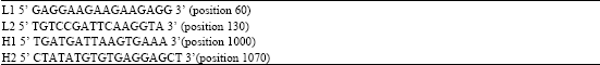
The c-myc gene exon 2 part (approximately 1.1 kb long) was amplified by using the Polymerase Chain Reaction (PCR) with these primers listed in Table 1.
The PCR amplification was carried out with specifically designed primers from the Genbank. In the primer design the trout, the canary, the fish and the frog myc gene exon 2 sequences were compared and because of the sharks more close to the fish then the others, in the fish (Cyprinus carpio) c-myc gene sequences, the similar parts were used in the primer design.
Perkin Elmer DNA Thermal Cycler 480 or a PTC-100TM Programmable Thermal Controller (MJ Research, Inc.) were used for all PCR amplifications. After extraction of tissue sample, the double stranded genomic DNA was used in a 30 cycles of PCR amplification. 5 min initial denaturing at 94°C, followed by 1 min at 94°C and either 1 min annealing at 50-55°C , followed by 2 min extension at 65°C or 3 min annealing plus extension at 60-65°C, after 30 cycle in final 10 min extension at 65°C. 50 μL reaction contains, 200 ng μL-1 of two external primers, 10 mM of each nucleotide (dATP, dCTP, dGTP and dTTP) ultrapure dNTP set from Pharmacia Biotech, 17.5 mM MgCl2 and 1.75 U of Taq polymerase (Boehringer Mannheim)or Tag polymerase (ExpandTM High Fidelity PCR system, Boehringer Mannheim) and at least 300 ng genomic DNA. After the first PCR, PCR sample was run on a 1% low melt agarose gel. Then the band, was visualised under low intensity ultraviolet light, excised and melted in a 65°C heating block. This excised amplified band amplified again with same PCR technique by using same or internal primers. The final fragments were purified for sequence by using high pure PCR product purification kit, according to kit instruction.
Cycle sequence reaction was carried out with using either a Perkin Elmer DNA Thermal Cycler 480 or a PTC-100TM Programmable Thermal Controller (MJ Research, Inc.).
| Table 1: | Primers used in PCR amplification and sequencing of c-myc gene exon 2. L- strand primer forward and H- strand for reverse primer |
 | |
The PCR amplified products were sequenced directly by using dideoxy chain termination method. 20 μl dye-termination reaction contains 300-500 ng of purified PCR amplified product, 5 pmol each primer and 20 mM MnSO4. The cycle includes first, denaturation at 96°C for 1 min, follow by 25 cycle of 50 sec at 96°C, 4 min at 62°C and 20 sec at 50°C. After that, the reactions were loaded onto ABI 373A automated sequencers.
Results
They are also unique animals in some cases that have some specific protective immune system against to the such as cancer. Up to know cancer never been recorded in sharks. Actually they have a cancer c-myc gene, but either is not active or have covered so that can’t be active. Despite number of attempt, the sequence was not obtained for that gene to determine bases code from different sharks. In all attempts, same level of pigment with a background for the sequence was obtained.
DISCUSSION
Because of not seeing cancer in sharks, the Genbank have been searched and found nothing about cancer-gene correlation in sharks in more general in cartilaginous fishes. Several papers have been found, suggesting a possible connection with a high level of urea in sharks, causing to immunisation against to the cancer or being made of cartilage again leads to the anticancer effects in elasmobranchs.
Sharks and rays have a very high level of intra and extracellular concentrations of urea because they use this urea as an osmotic adaptation for life in salt water that happens also some other primitive fishes. The important meaning for that, urea can disrupt DNA transcription and form inactive proteins which have mostly found in marine elasmobranchs, that means molecular evolution in this group is occurring in an extreme cellular environment and also this may effect the evolution of protein, for that reason, may be sharks seems to have higher amino acid sequence conservation than the mammals. Furthermore, in marine sharks, some proteins need urea for normal function, by using some specific amino acids for adaptation to cellular stress. If you compare rates and patterns of amino acid replacement between shark and mammals, amino acid replacement rates in sharks are found approximately 6 times slower than in mammals by sequencing mitochondrial cytochrome b gene (Martin and Palumbi, 1993). The much slower rates of the amino acid replacements or in other words much higher amino acid conservation in sharks indicates mutation or turning to the cancer cells in sharks occurs very rarely. This result was supported with no cancer records in sharks. If you look at different paper, they all give different approach to the question of the why never been found or seen or reported cancer in sharks. Different shark-related material and products were tested and suggested in the explanation of the shark- cancer free situation, creating a link.
For example, the rarity of tumors in elasmobranchs is because of the high ionic strength in their tissue, causing to a high body temperature, were suggested by Rosen and Woodhead (1980). It is known that, sharks’ body temperature 7-10°C higher than the water temperature, because heat was kept inside of the body via using the heat exchangers through the circulatory system. This unique environment should lead to the elimination of variable antigenicity and consequently100% immune surveillance. If the repressor molecular of mitosis doesn’t bind to its operator site, can not bind anywhere, reality was suggested for this explanation (Rosen and Woodhead, 1980). In mitosis the role of the iğ flaments can be disturbed by some alkaloids, such as; colchisin blocks iğ flaments’ protein formation and chromatide separation while in nucleus dividing. Consequently cell and nucleus dividing can’t be complete. These antimitotic characteristics provide using them as onkolitic (anticancer) materials in cancer cases.
In elasmobranch fishes, urea occurs at high concentrations (350-600 mM) in the body fluids and tissues, where it plays an important role in osmoregulation. Because of not seeing cancer cases in elasmobranchs reminds a possible use of the urea in cancer cases. A series of N-aryl-N'-(2-chloroethyl) urea (CEUs) and derivatives were tested for antiproliferative activity against a wide panel of tumor cell lines. The immunofluorescence microscopy assays, indicates that these compounds does some changes in the cytoskeleton and but don’t give any damage to DNA. Because of this result, CEUs are accepted as a new class of antimitotic agents (Mounetou et al., 2001). Comprising cartilage in cartilaginous skeleton instead of bone, made elasmobranchs different compare to the other animals. This unique structure can be effective in elasmobranchs’ immunity against to cancer. Because of these, in finding of the effective cancer treatment, shark cartilage and derivatives and shark liver oil also were examined as possibly new anticancer materials.
Shark cartilage include a substance that prevents the growth of new blood vessels toward solid tumors, hence tumor growth fails. Being high amount of this factor in shark cartilage, may make sharks an ideal source in using against to tumor formation and therefor the rarity of neoplasms in these animals can be explained (Lee and Langer, 1983). The use of crude cartilage for the treatment of human cancers remains controversial. The properties of AE-941, a novel standardised water-soluble extract derived from shark cartilage that represents less than 5% of the crude cartilage was tested. This compound includes several biologically active molecules, works as multifunctional antiangiogenic product. The effectiveness of this compound against to different cancer type was searched in Europe and North America. A potent angiogenesis inhibitor, U-995, has been extracted from the blue shark (Prionace glauca) cartilage. The role of the U-995, as inhibitor against to tumor cell growth and metastasis, was determined (Sheu et al., 1998).
Another suggestion using as an inhibitory product of shark serum against to Lewis lung carcinoma was examined in C57BL/6J mice. After an ip injection of Carcharhinus plumbeus, the sandbar shark’s whole serum into this mice, infected with Lewis lung carcinoma, tumors failed to develop in 69% on days 0, 3, and 6 after tumor transplantation (Snodgrass et al., 1976).
Shark has a very good source of liver oil with including almost 10 times higher amount of liver oil compare to the other animals. Shark liver oil has been used for over 40 years with a therapeutic and preventive agent purposes. The effective parts of shark liver oil have been determined as a group of ether-linked glycerols known as alkylglycerols. Studies have been going on over the last 30 years, indicates that alkylglycerols have a multifunctional role. The natural alkylglycerols level goes higher within tumor cells, possibly to control cell growth. The activation of protein kinase C, an important step in cell proliferation, can be prevented by alkylglycerols. The alkylglycerols can be obtained easily from shark liver and can be used up to 100 mg three times a day dosage with no known side effects in the infectious diseases treatment (Pugliese et al., 1998).
Since angiogenesis is a key feature of tumor growth, inhibiting this process is one way to treat cancer. Cartilage is a natural source of material with strong antiangiogenic activity. Because sharks’ endoskeleton was made of cartilage, sharks are thought to be an ideal source of angiogenic and tumor growth inhibitors. Shark cartilage extract has shown antiangiogenic and antitumor activities in animals and humans. Purified antiangiogenic factors from shark cartilage, such as U-995 and neovastat (AE-941), also showed antiangiogenic and antitumor activity (Cho and Kim, 2002).
Bone metastases are generally associated with bone destruction which occurs in response to factors secreted by metastatic cells. In the determination of Neovastat (AE-941), as a multi-functional inhibitor of angiogenesis, Neovastat (AE-941) was used in the analysis and radiographic analysis indicated that Neovastat decreased the number of osteolytic lesions by 33% (p≤0.3). Moreover, a decrease in the tumor volume in bone was observed in Neovastat-treated animals. These results indicate that Neovastat may be useful in preventing bone metastasis in cancer patients (Weber et al., 2002).
Neovastat (AE-941) is an antiangiogenic drug isolated from marine cartilage. It interferes with several steps associated with the development of angiogenesis via its ability to induce endothelial cell apoptosis, and inhibiting the some signaling pathways, suggesting, Neovastat behaves as a multifunctional antiangiogenic drug. Neovastat shows significant antitumor and antimetastatic properties in animal models. This indicates that Neovastat is suitable for long-term use, either alone or in combination with other anticancer therapies (Gingras et al., 2003).
CONCLUSIONS
Different materials, related to elasmobranchs in more specifically sharks, were examined and suggested in the treatment of the tumor, cancer cases. The important point is that, all of them indicates something unique to the sharks, effective on immunisation against to tumor formation and prevents sharks from cancer. This explanation also is supported by the reality of not seen or recorded any cancer cases in sharks. But still needs to be checked in detail to tell more on this subject. Roughly one person in five die because of the cancer. Some other problems such as malnutrition, heart disease and stress are may be more serious but cancer has a unique situation by disturbing the fundamental rules of the cell behaviour. A lot of massive researches are undergoing in cancer treatment and obtained very important benefits from the most of them, but still the certain treatment or cure is far from the applying into the practice.
REFERENCES
- Cho, J. and Y. Kim, 2002. Sharks: A potential source of antiangiogenic factors and tumor treatments. Mar. Biotechnol. 4: 521-525.
CrossRef - Gingras, D., D. Boivin, C. Deckers, S. Gendron, C. Barthomeuf and R. Beliveau, 2003. Neovastat-a novel antiangiogenic drug for cancer therapy. Anticancer Drugs, 14: 91-96.
PubMedDirect Link - Mounetou, E., J. legault, J. Lacroix and R. Gaudreault, 2001. Antimitotic antitumor agents: Synthesis, structure relationships and biological characterization of N (2-chloroethyl) ureas as new selective alkylating agent. J. Med. Chem., 44: 694-702.
Direct Link