
S. Mansouri
Department of Microbiology, Kerman University of Medical Sciences, P.O. Box 444, Kerman, Iran
E. Ghasami
Department of Microbiology, Kerman University of Medical Sciences, P.O. Box 444, Kerman, Iran
N. Shahabi Najad
Koladuz (Hazraet Fatme) Hospital, Shariaeti Street, Kerman, Iran
Journal of Medical Sciences
Year: 2008 | Volume: 8 | Issue: 6 | Page No.: 574-578


ABSTRACT
The objective of present study was to determine reliable data on vaginal carriage, serotype distribution and antibacterial susceptibility of Streptococcus agalactiae, Group B streptococcus (GBS) in pregnant women in southeast of Iran. Vaginal swab cultures for GBS were obtained from 602 pregnant women at childbirth. Susceptibility of the isolates to penicillin, ampicillin, clindamycin and erythromycin were determined by standard agar dilution method. Isolates were classified according to their capsular polysaccharide types. GBS was isolated from 55 pregnant women (9.1%). All isolates were sensitive to penicillin and ampicillin [Minimum Inhibitory concentration (MIC) range of 0.03-16 μg mL-1 and ≤ 0.03-1 μg mL-1, respectively). Erythromycin and clindamycin resistance were seen in 10.9% (MIC range ≤ 0.03-16 μg mL-1) and 25.4% (MIC range ≤ 0.03-32 μg mL-1) of the isolates, respectively. Serotype III (41.8%), Ib (25.45%) and II were the most frequently isolated serotypes (14.54%). Group IV was not detected and 14.54% of the isolates were non type-able. No correlation was found between GBS colonization and demographic factors of age, parity, history of abortion or ruptured membrane and vaginal signs or symptoms. In conclusion the rate of GBS colonization is low in this area, but serotype III, which is mostly involved in invasive disease is the predominant serotype. Routine maternal screening should be performed to prevent group B streptococcal disease in neonates in this district. Susceptibility to the isolates to clindamycin and erythromycin should be checked in the penicillin-allergic patients, to avoid treatment failure.
PDF Abstract XML References Citation
How to cite this article
S. Mansouri, E. Ghasami and N. Shahabi Najad, 2008. Vaginal Colonization of Group B Streptococci During Late Pregnancy in Southeast of Iran: Incidence, Serotype Distribution and Susceptibility to Antibiotics. Journal of Medical Sciences, 8: 574-578.
DOI: 10.3923/jms.2008.574.578
URL: https://scialert.net/abstract/?doi=jms.2008.574.578
DOI: 10.3923/jms.2008.574.578
URL: https://scialert.net/abstract/?doi=jms.2008.574.578
INTRODUCTION
Streptococcus agalactiae or Streptococcus group B (GBS) is an important pathogen causing various infections in neonates and pregnant women and opportunistic infections in immunocompromised patients and adults with serious infections (Efstratiou et al., 2006). Although, protocols for the prophylaxis and screening of pregnant women before delivery is recommended in many countries, GBS is still the leading cause of early onset sepsis in the neonates and an important cause of neonatal morbidity and mortality worldwide (Goldenberg and Thompson, 2003; Baron, 2003). Women with vaginal, rectal or Urinary tract colonization of GBS can vertically transmitted the bacteria to their newborns (El-Kersh et al., 2002). The vaginal colonization rate of GBS in reported to be between 5-40% around the world and colonization can be transient, chronic or intermittent (El-Kersh et al., 2002; Amin et al., 2002; Barcaite et al., 2008; Chohan et al., 2006; Motlova et al., 2004). The routine screening of pregnant women at the 35-37 weeks pregnancy and subsequent prophylaxis for women known to be colonized with GBS or those evaluated to be at high risk, is recommended by many countries and had been shown to prevent many causes of early onset diseases in the babies (Schrag et al., 2002). Penicillin G is the drug of choice for the treatment of GBS and there is no report on GBS resistance to penicillin. Erythromycin and clindamycin are alternative antibiotics recommended for the intrapartum prophylaxis in penicillin allergic GBS carriers (Efstratiou et al., 2006; Baron, 2003).
Resistances of GBS isolates to antibacterial agents except penicillin and ampicillin are reported to be increasing (Barcaite et al., 2008; Uh et al., 2004; Zeng et al., 2006; Dela Cruz et al., 2007; Dogan et al., 2005) and therefore, regional surveillance for the antibiotic resistance is recommended. Another way to prevent the early onset disease in the newborn is to immunize the high risk mothers at the time of delivery. The distribution of different GBS serotypes in different parts of the word may not be the same, which makes difficulty in vaccine preparation (Efstratiou et al., 2006; Baron 2003; Uh et al., 2004; Ko et al., 2001). To prepare an accurate vaccine in a region, a population based serotype survey is needed to be insuring about the proper mix of the serotypes in the vaccine preparations. Presently there are nine major serotypes of GBS with a few serotypes that are more involved in the human infections (Efstratiou et al., 2006; Dogan et al., 2005; Ekin and Gurturk, 2006). Although, in several studies the GBS colonization rate in various part of Iran is determined, this is the first report on colonization rate, sensitivity of the isolates to the first line treatment options and the serotype distribution of GBS isolates in pregnant women in the southeast of Iran.
MATERIALS AND METHODS
Vaginal samples (602) were collected from pregnant women attending the three major non-private hospitals in the southeast of Iran (Kerman), during April 2006 to March 2007. The samples were collected at 35 to 37 weeks of gestation or at delivery from women who agreed to participate in the study. A history sheet was completed according to the information obtained from the patients with the variables shown in Table 1. Women with the fever, ruptured membrane or those who received antibiotics in the two weeks prior to sampling were excluded from the study. The swabs were inoculated into Amies transport media and were transported to the laboratory within 24 h. The swabs were cultured into Todd-Hewitt broth for enrichment. Subcultures were made on the 5% sheep blood agar plates containing 8 μg mL-1 gentamicin and 15 μg mL-1 nalidixic acid to prevent the growth of normal bacterial flora (El-Kersh et al., 2002). The β-hemolytic gram positive catalase negative isolates were identified as GBS by conventional methods (MacFaddin, 2000). The isolated GBS were kept at Cryo Bank (Mast Co England) at -70 °C for further analysis.
Antibacterial resistance against amicillin, erythromycin, clindamycin and penicillin were determined by standard methods of agar dilution (Forbes et al., 2007). Serotyping was performed on the GBS isolates using hemolytic streptococci group B typing sera from Mast (England). The commonly isolated serotype l, la, lb, ll, III, V and lV were used.
The data were analyzed using SPSS version 11 (SPSS Inc., Chicago, 111). The Fisher test was used for categorical data. p-values of ≤ 0.05 was considered significant (two-tailed test).
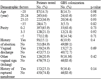
| Table 1: | Relation between GBS colonization and demographic factors in pregnant women in southeast of Iran |
 | |
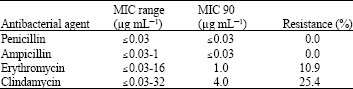
| Table 2: | Antimicrobial susceptibility of group B streptococci isolates (n = 55) from colonized pregnant women in the southeast of Iran |
 | |
RESULTS
From a total of 602 vaginal swabs from pregnant women 55 (9.1%) isolates were identified as the group B streptococci (GBS). There was no significant difference in the rate of vaginal GBS colonization among pregnant women in respect to age, parity, history of abortion and history of membrane rupture, or vaginal sign and symptoms (Table 1).
All of the 55 GBS isolates were found to be sensitive to penicillin and ampicillin. Sensitivity to erythromycin was also high and 49 isolates (89.1%) were sensitive to this agent (Table 2). Resistance to clindamcin was found in 14 (25.4%) of the isolates and the MIC range of the resistant strains was 4-32 μg mL-1 (Table 2). Simultaneous resistance to both clindamycin and erythromycin was found in two isolate.
By using specific antisera against commonly isolated GBS, 47 GBS serotypes were isolated, the serotypes in order of decreasing frequencies were: serotype III (23 isolates, 41.8%), serotype Ib (14 isolate, 24.45%), serotype II (8 isolate, 14.54%), serotype la (1 isolates, 1.8%) and serotype V (1 isolates, 1.8%). Serotype IV was not detected and 8 isolates were not typeable with the used antiserums.
DISCUSSION
The carriage rate of Streptococcus agalactiae, (Streptococcus group B, GBS) in southeast of Iran was determined to be 9.1% in this study. This is in agreement with other investigations in Iran with the rate of 9.2% reported from one center in Kerman and 8% from Tehran, respectively (Aali et al., 2007; Bakhtiari et al., 2007). However the rate of 5.2% for GBS colonization has been found in northwest of Iran which is lower than our findings (Nahaei et al., 2007). No correlation was found between GBS colonization rate and the demographic factors shown in the Table 1. The results obtained by GBS association and demographic factors are conflicting. For example a significantly higher rate of colonization with a history of spontaneous rupture of membrane and vaginal colonization was reported by El-Kersh et al. (2002). A correlation with age and GBS carriage is reported by Aali et al. (2007) but was not seen by Schrag et al. (2002) and Honig et al. (2002). Since GBS carriage is usually reported to be asymptomatic (Goldenberg and Thompson, 2003) and the results on demographic factors are not all the same, identification of risk factors for colonization requires further studies.
Antibacterial resistance pattern of the isolates to four common therapeutic agents used for GBS prophylaxis showed no resistance to penicillin and ampicillin; however two isolates had elevated level of MIC against ampicillin (1 μg mL-1) which is not in the resistance category. This result confirms the uniform susceptibility of GBS isolates to the mentioned drugs (Efstratiou et al., 2006; Baron, 2003; Motlova et al., 2004; Zeng et al., 2006). Resistance to erythromycin and clindamycins which are alternative drugs in the patients allergic to beta-lactams, are increasing in the GBS isolates (Chohan et al., 2006; Uh et al., 2004). Erythromycin resistance in western countries is reported to be in a range of 4 to 26.9% (Chohan et al., 2006; Dogan et al., 2005; Berkowitz et al., 1990; Savoia et al., 2008), while, resistance to clindamycin is about the same or lower. The GBS isolates from Korea and Taiwan had higher sensitivity to erythromycin compared to clindamycin (Uh et al., 2004; Ko et al., 2001). However, in this study resistance to erythromycin and clindamycin were 10.9 and 25.4%, respectively, with two isolates were resistance to both agents simultaneously. The most common serotypes isolated in this study were serotype III (41.8) and Ib (25.45%). In accordance with present findings, serotype III is reported to be the most common GBS serotype around the world (Barcaite et al., 2008; Motlova et al., 2004; Uh et al., 2004; Savoia et al., 2008). In contrary in the northwest of Iran (Tabriz), serotype V is reported to be the prevalent serotype (19.5%) and the serotypes III and IV had the lowest frequency (9.5 and 8.2%), respectively (Nahaei et al., 2007). Non type-able GBS in present study were 15.45%, which is similar to the results obtained in Tabriz, United Arab Emirates and Korea (Amin et al., 2002; Uh et al., 2004; Nahaei et al., 2007). Some of non-type able strains could be due to uncommon serotypes (Ic, VII and VIII) which were not tested in this study. A relation between serotype and antibacterial resistance in GBS isolates is reported by Dogan et al. (2005) especially erythromycin resistance and serotype V. Savoia et al. (2008) reported the higher rate of resistance to erythromycin in the serotypes that are more prevalent in Italy (III, V and Ia). In this study although a higher rate of resistance to erythromycin and clindamycin was found in the serotype III and V the difference were not significant.
The colonization rate of GBS in this region was not high, but the majority of isolates belonged to serotype III, which is mostly involved in neonatal infections in human.
Many risk factors for GBS colonization are still unknown and further study with a higher sample size is required to determine the risk factors for GBS colonization in pregnant women. Therefore, maternal screening strategy and proper prophylaxis or treatment should be more effective than the risk-based approaches for preventing group B Streptococcal disease in neonates in this district. Due to the emergence of clindamycin and erythromycin resistance in the GBS isolates, the isolates should be tested for the susceptibility to these drugs in the penicillin-allergic patients.
ACKNOWLEDGMENTS
The authors are grateful to the doctors and midwives in the departments of obstetrics and gynecology of the Afzalipoor, Khashani and Kolahdooz hospitals in Kerman, for their help in collection of samples. This study was supported by a grant No. 83/27 from Research Council of Kerman University of Medical Sciences.
REFERENCES
- Aali, B.S., H. Abdollahi, N. Nakhaee, Z. Davazdahemami and A. Mehdizadeh, 2007. The association of preterm labor with vaginal colonization of group B streptococci. Iranian J. Reprod. Med., 5: 191-194.
Direct Link - Bakhtiari, B., M.M. Soltan Dallal, M.J. Zaeim Yazdi, J. Fallah and M.N. Amir et al., 2007. Evaluation of PCR method for diagnosis of group B Streptococcus carriage in pregnant women. Iran. J. Med. Microbiol., 1: 1-8.
Direct Link - Barcaite, E., A. Bartusevicius, R. Tameliene, M. Kliucinskas and L. Maleckiene et al., 2008. Prevalence of maternal group B Streptococcal colonisation in European countries. Acta. Obstet. Gynecol. Scand., 87: 260-271.
CrossRef - Baron, E.J., 2003. Laboratory support for prevention of perinatal group B streptococcal disease: Commentary on the new guidelines on screening for group B Streptococci during pregnancy. Clin. Microbiol. Newsletter, 25: 65-69.
CrossRef - Berkowitz, K., J.A. Regan and E. Greenberg, 1990. Antibiotic resistance patterns of group B streptococci in pregnant women. J. Clin. Microbiol., 28: 5-7.
PubMedDirect Link - Chohan, L., L.M. Hollier, K. Bishop and C.C. Kilpatrick, 2006. Patterns of antibiotic resistance among group B Streptococcus isolates: 2001-2004. Infect. Dis. Obstet. Gynecol., 2006: 57492-57492.
Direct Link - Dela Cruz, W.P., J.Y. Richardson, J.M. Broestler, J.A. Thornton and P.J. Danaher, 2007. Rapid determination of macrolide and lincosamide resistance in group B streptococcus isolated from vaginal-rectal swabs. Infect. Dis. Obstet. Gynecol., 2007: 46581-46581.
Direct Link - El-Kersh, T.A., L.A. Al-Nuaim, T.A. Kharfy, F.J. Al-Shammary and S.S. Al-Saleh et al., 2002. Detection of genital colonization of group B streptococci during late pregnancy. Saudi Med. J., 23: 56-61.
Direct Link - Goldenberg, R.L. and C. Thompson, 2003. The infectious origins of stillbirth. Am. J. Obstet. Gynecol., 189: 861-873.
PubMed - Honig, E., J.W. Mouton and W.I. Van der Meijden, 2002. The epidemiology of vaginal colonisation with group B streptococci in a sexually transmitted disease clinic. Eur. J. Obstet. Gynecol. Reprod. Biol., 105: 177-180.
Direct Link - Ko, W.C., H.C. Lee, L.R. Wang, C.T. Lee and A.J. Liu et al., 2001. Serotyping and antimicrobial susceptibility of group b streptococcus over an eight-year period in Southern Taiwan. Eur. J. Clin. Microbiol. Infect. Dis., 20: 334-339.
CrossRef - MacFaddin, J.F., 2000. Biochemical Tests for Identification of Medical Bacteria. 3rd Edn., Lippincott Williams & Wilkins, Pennsylvania, United States, ISBN: 0-683-05318-3, Pages: 912.
Direct Link - Motlova, J., L. Strakova, P. Urbaskova, P. Sak and T. Sever, 2004. Vaginal and rectal carriage of Streptococcus agalactiae in the Czech Republic: Incidence, serotypes distribution and susceptibility to antibiotics. Indian J. Med. Res., 119: 84-87.
Direct Link - Nahaei, M.R., N. Ghandchilar, N. Bilan and P. Ghahramani, 2007. Maternal carriage and neonatal colonization of streptococcus agalactiae in Tabriz, Northwest Iran. Iranian J. Med. Sci., 3: 177-181.
Direct Link - Savoia, D., C. Gottimer, C. Crocilla and M. Zucca, 2008. Streptococcus agalactiae in pregnant women: Phenotypic and genotypic characters. J. Infect., 56: 120-125.
CrossRefPubMedDirect Link - Schrag, S.J., E.R. Zell, R. Lynfield, A. Roome and K.E. Arnold et al., 2002. A population-based comparison of strategies to prevent early-onset group B streptococcal disease in neonates. N. Engl. J. Med., 347: 233-239.
Direct Link - Zeng, X., F. Kong, H. Wang, A. Darbar and G.L. Gilbert, 2006. Simultaneous detection of nine antibiotic resistance-related genes in Streptococcus agalactiae using multiplex PCR and reverse line blot hybridization assay. Antimicrob Agents Chemother., 50: 204-209.
Direct Link