
I.C . Mehta
Department of Computer Technology, MIET, Gondia, India
Z.J . Khan
RCERT, Chandrapur, India
R.R . Khotpal
LIT, Nagpur, India
Information Technology Journal
Year: 2006 | Volume: 5 | Issue: 3 | Page No.: 577-582


ABSTRACT
Chest radiograph inherently display wide dynamic range of X ray intensities. A wide variety of preprocessing procedures for chest radiograph based on local equalization, sharpening, fuzzy set, neural network and combination of modification of these techniques have been proposed. In this paper we have made analysis of twelve different techniques specifically derived and tested for enhancement of chest X ray giving brief details of working of algorithm. Radiology department are staring use central system to store digital chest radiograph. This provides opportunity to use chest workstation with different enhancement algorithm that run in background and radiologist may view a patient radiograph using different enhancement algorithm for subsequent analysis. Enhancement algorithm should be selected based on radiologist requirement and disease specific. Hence it is necessary to make review and analysis of available chest radiograph enhancement technique.
PDF Abstract XML References Citation
How to cite this article
I.C . Mehta, Z.J . Khan and R.R . Khotpal, 2006. Analysis and Review of Chest Radiograph Enhancement Techniques. Information Technology Journal, 5: 577-582.
DOI: 10.3923/itj.2006.577.582
URL: https://scialert.net/abstract/?doi=itj.2006.577.582
DOI: 10.3923/itj.2006.577.582
URL: https://scialert.net/abstract/?doi=itj.2006.577.582
INTRODUCTION
In medical applications, image contrast enhancement is important for the diagnosis of many diseases. In chest radiography the image contrast is low due to less difference in X ray attenuation coefficients. The development of enhancement is based on visual principles. The human eye is sensitive to high frequency signals, their visibility become low when they are embedded in high frequency background signals. The proper amplification of the high frequency components in medical image will improve visual perception and help the diagnosis. One of the most important difficulties in image enhancement is quantifying the chest radiograph criteria for enhancement. One of the most challenging tasks in diagnostic radiology is to generate clinically useful images with improved visualization for diagnosis. In the present investigation enhancement technique are useful for obtaining better visibility, clinical information such as pulmonary infiltrates, heart size, rib lesion, pleural effusion, pneumothorax, lung nodules etc.
Chest radiograph inherently display wide range of x ray intensities. In unprocessed X ray it is often hard to >see through= the mediastinum and contrast in the lung field is limited. A classical solution to this kind of problem in image processing is use of (local) histogram equalization technique. A related technique is enhancement of high frequency details (sharpening). Such techniques are so essential for optimized display of image in soft-copy reading. Enhancement techniques based on various combinations of methods are possible. For chest radiograph crisp and fuzzy based enhancement technique have been discussed.
REVIEW OF CHEST RADIOGRAPH ENHANCEMENT
Method: We have taken chest radiograph from the various radiologist and obtained clinical diagnosis from the two radiologist. We have applied various algorithm for the enhancement of chest radiograph given by various authors and compared results with the help of radiologist at computer center of MIET. Final conclusion was drawn by taking into account the author and radiologist comments.
Neuro equalizer: The Neuro-equalizer has multi layer structure with sigmoid activation function and use error back-propagation algorithm. The neural network has two inputs. An input to neural network is mean value of images pixels and another input is gray level of pixel at chest image. The output of neural network is gray level of improved pixel that is equalized gray level of the pixel
Region Based Enhancement (RBE): The algorithms utilize wavelet-filtering technique to enhance the image features of selected anatomic region of interest in chest radiograph. Wavelet filtering is applied to original image and two images are obtained one containing mostly low frequency components and other high frequency components. The low passed image provides the information of where are the gray regions of interest. The high pass contains high frequency components such as the vascular structure, which are to be enhanced. The result shows enhanced visibility of the spine and vascular structure in medistinum area without degrading the contrast and intensity of the lung fields (Shih-Chung and Jhy-Shyam, 1996).
Just Noticeable Difference Guided Adaptive Contrast Enhancement (JGACE): The effective contrast enhancement diagnostic purpose of chest radiograph can be achieved by including certain basic human propertied. This is a novel adaptive algorithm that tailors the required amount of contrast enhancement on local contrast of the image and the observers Just Noticeable difference. The schematic diagram of JGADE algorithm is and consists of Low pass filtering, Separating Details and Smooth Regions, Determination of Local Spatial Frequency and Contrast, Determination of Local Contrast Gain, The Transformation F (.) (Ji and Sundareshan, 1996).
Regionally Adaptive Histogram Equalization (RAHE): Histogram equalization is a well-known technique where image quality is improved by equally distributing pixel intensity throughout the available gray scale (Hummel, 1977; Castelman, 1979). This is accomplished by creating a look-up-table gray-scale transformation based on the cumulative distribution function. For chest images it is better to perform local area histogram equalization, thereby improving contrast in these smaller regions (Johnson et al., 1985). In order to apply this adaptive histogram equalization scheme to digital chest radiographs several factors need to be considered. Structures in the mediastinum and subdiaphragm need to be enhanced selectively.
The transition from the processed mediastinum to the unprocessed lungs must be smooth and free of artifacts that the radiologist might misinterpret.
Enhancement Based on Visual Model Using the Concept of Fuzzy Set (EVMFS): For enhancing the contrast of the chest radiograph the fuzzy based enhancement technique discussed is used by Shen and Wan (1992), Johnson et al. (2000) use enhancing the contrast of the chest radiograph the technique discussed. An image X of M*N dimension of L level can be considered as an array of fuzzy singleton with a value of membership function and indicating the degree of brightness level I = 0 1 2..L-1. It is possible to transform the spatial plane to fuzzy property plane with the help of an asymmetry S-function. For obtaining the desired amount of enhancement there are many operator, which can be used for different area of enhancement application (Hariharan et al., 2000). These operators on fuzzy set generate another fuzzy set.
Automatic Anatomically Selective Image Enhancement (AASIE): Automatically selective enhancement is motivated by the desire to simultaneously meet the different enhancement requirement of the lung field and medistinum. A peak detection algorithm and set of rules are applied to the image histogram to determine automatically a gray level threshold between the lungs filed and medistinum. The gray level threshold facilitates anatomically selective gray-scale modification and/or unsharp masking. Further, in attempt to suppress possible white band or black-band artifacts due to unsharp masking at sharp edges, local contrast adaptivity is incorporated into anatomically selective unsharp masking by designing an anatomy sensitive emphasis parameter which varies as asymmetrically with positive and negative values of the local image contrast (Johnson et al., 1985; Kundel et al., 1979).
Chest Radiograph Image Enhancement by Adaptive Processing (IHAP): The algorithms discussed here uses the knowledge about the shape and contrast present in original image by the structure in specific the lung nodules in order to enhance them adequately without excessively affecting the large amount of high frequency information found in lung field, thus improving the diagnostic capacity. For each pixel, the algorithms calculates a local contrast on the original image using a neighborhood criteria adapted to the information of its surrounding and modified it using a contrast enhancement function (cef) the design of which depends on the region of the contrast histogram we wish to enhance. The final image is obtained by an inverse transformation in the enhanced contrast domain to the image domain (Sherrier and Johnson, 1987; Sezan et al., 1989; Carreira et al., 1991).
Enhancement Using Automatic Spatial Filtering Technique (EASFT): Processing consists of following steps (McNitt-Gray et al., 1991).
| • | Obtain two smoothed images from original image. |
| • | Build a linear combination of these three images. |
| • | Calculate a nonlinear constant function transform. |
| • | Nonlinear constant stretching with transform generated on the third step. |
Adaptive Chest Radiograph Enhancement Using First Derivative Operator and Local Statistics (AEFDLS): The algorithm consists of three processing steps (Sherrier and Johnson, 1987).
| • | Remove film artifacts |
| • | Compute the gradient image by using the first derivative operator. |
| • | Enhance the important features of the radiographic image by adding the adaptively weighted gradient image. In this local statistics of the image is utilized for the realization of the enhancement, so that image details can be enhanced and noise can be suppressed |
Fuzzy Method of Contrast Enhancement (FCE): A simple rule based approach to contrast enhancement can be formulated (Krell et al., 1997; Kam and Hanmandly, 2003)
| • | Setting the parameter of inference system (input features, membership functions) |
| • | Fuzzification of the actual pixel (membership to the dark, gray and bright sets of pixels) |
| • | Inference (if dark then darker, if gray then gray, if bright then brighter) |
Defuzzification of the interference results by the use of three singletons.
Nonadaptive Unsharp Masking (NUM): Historically, the nonadaptive unsharp masking technique was first implemented photographically (Levi, 1974). In the photographic unsharp masking process, the print obtainable from a negative was improved by preparing a blurred positive and printing the two in register. Digital unsharp masking follows the same principle. First, an unsharp mask is computed by low-pass filtering the original image. This is subtracted from the original to obtain a high-frequency image. Then the high-frequency image is amplified by an emphasis parameter and added back to the original image to yield the enhanced image (Ibrahim Sezan et al., 1989). Adaptive Unsharp Masking (AUM) Adaptive unsharp masking is achieved when the value of the high-frequency emphasis parameter is varied with some local property of the image, such as the pixel value and/or local contrast. In anatomically selective unsharp masking, the emphasis parameter varies with the pixel value. Further, the lung/mediastinum gray-level threshold is incorporated into the design to make it anatomy sensitive (Lim, 1984).
DISCUSSION
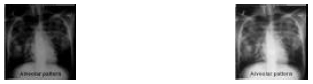
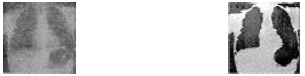
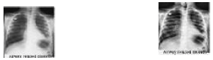
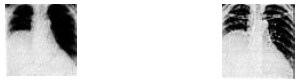
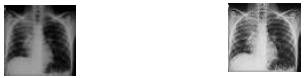
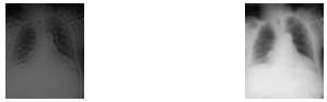
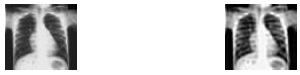
NE method has the good quality that radiologist observed. RBE algorithm can be applied to posterior-anterior and lateral chest radiograph in either vertical or horizontal orientation (Fig. 1). The artifact along the lung field boundary can be reduced by suppressing the high frequency component along the lung field location. JGACE brought up an adequate contrast for the low-contrast sinusoidal pattern without ringing artifacts. The background noise was not enhancement in uniform region. It was observed that high contrast signals in the chest radiograph image are kept unchanged by JGACE because their contrast is judged by algorithm on the basis of normal visual just noticeable difference. In RAHE the histogram of mediastinum is narrower in sub diaphragmatic regions; the histogram range is very limited (Fig. 2). All these functions are used to direct the processing of chest radiograph, enhancing substantially contrast of region of interest. With this technique histogram are calculated locally and modified according to both the mean pixel value of that region as well as certain characteristics of the cumulative distribution function. In EVMFS the image is first segmented using fuzzy Kohonan neural network and enhancement technique utilizes the updated fuzzy membership of an asymmetric function (Fig. 3). The result show well defined boundaries with enhancement that can provide several clues to the physician and radiologist for their clinical diagnosis. AASIE is based on auto threshold selection technique based on peak detection algorithm to determine lung/medistinum gray level threshold using histogram of chest radiograph (Fig. 4). The threshold is then used to implement anatomically selective gray scale transformation and unsharp masking. IHAP algorithms uses knowledge about shape and contrast presented in the original image and enhances in particular lung nodules without excessively affecting the large amount of high frequency information found in lung field (Fig. 5). It was observed that pathology were recognizable in the processed image. EASFT algorithm enhances the chest radiograph while minimally amplifying noise and provides better-enhanced output the histogram equalization. AEFDLS algorithm highlights lung boundaries and edges of the bones showing the contour of the lung clearly. FCE algorithm enhances contrast in the image keeping the original characteristics of the image unchanged (Fig. 6). This algorithm makes better visibility of inter rib regions. In NUM image enhancement is based on preparing a blurred positive version of the negative and superimposing it on the negative (Fig. 7). Range of frequency to be enhanced depends on size of local window. In AUM the emphasis parameter can be varied with respect to the local image contract in an attempt to suppress possible gray-level overshoots and undershoots (dark-band and white-band artifacts) due to high local image contrast at the sharp edges (Fig. 8).
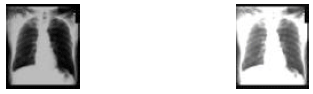 | |
| Fig. 1: | The original image and enhanced image based on RBE |
 | |
| Fig. 2: | The original image and enhanced image based on RAHE |
 | |
| Fig. 3: | The original image and enhanced image based on EVMFS |
 | |
| Fig. 4: | The original image and enhanced image based on AASIE |
 | |
| Fig. 5: | The original and enhanced image based on IHAP |
 | |
| Fig. 6: | Original image after contrast-enhanced image based on FCE |
 | |
| Fig. 7: | The original image and enhanced image based on NUM |
 | |
| Fig. 8: | The original image and enhanced image based on AUM |
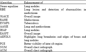
| Table 1: | Algorithms and specific enhancement of chest radiograph |
 | |
Computer aided analysis and interpretation of x-ray images can provide several useful clues for diagnosis, treatment planning and medical research. (Garland, 1989; Duncan and Ayache, 2000; Doi et al., 1999; Ginneken, 2001). Various chest radiograph enhancement techniques has been discussed in brief and analysis has been made.
ANALYSIS
Enhancement algorithm should be selected based on radiologist requirement and disease specific. Neuro equalizer enhances lung nodules. RBE enhances lung lesion and detection of abnormalities in medistinum It also enhances mediastinum and diaphragm with minimum change of lung field on the computed chest radiograph (Table 1). JGACE enhances overall image and image quality is rated as excellent by radiologist from diagnosis point of view. RAHE enhances regions such as mediastinum and subdiaphagmatic region are enhanced substantially. EVMFS enhances the chest radiograph images of TB patients for better diagnosis of diseases. AASIE enhances lung and mediastinum. IHAP enhances lung nodules in particular. EASFT enhances overall image. AEFDLS highlights lung boundaries and edges of bones and lung nodules. This algorithm enhances the lung nodules if any in chest radiograph and hence it has got clinical importance. FCE better visibility of inter rib region. Any abnormalities such as TB, lung nodules they can identified from the enhanced image. NUM and AUM enhances overall chest radiograph image (Table 1).
Enhancement algorithm should be selected based on radiologist requirement and disease specific
CONCLUSIONS
Researchers have proposed various algorithms for the overall and specific part of chest radiograph enhancement. Good enhancement algorithm for tuberculosis, distinguishing true nodule from (overlapping) shadow from vessels and ribs, benign or malignant lung nodule etc are to be further specifically developed. What is the need of the hour is single computer aided method which will enhance chest radiograph disease specific. Based on this analysis one can select the required enhancement techniques specific to the disease.
REFERENCES
- Carreira, M.J., D. Cabello A. Mosquera, M.G. Penedo and I. Facio, 1991. Chest X ray image enhancement by adaptive processing. Proceedings of the IEEE Annual International Conference on Engineering in Medicine and Biology Society, Oct. 31-Nov. 3, IEEE Xplore, pp: 1056-1057.
Direct Link - Duncan, J.S. and N. Ayache, 2000. Medical image analysis: Progress over two decades and the challenges ahead. IEEE. Trans. Pattern Anal. Mach. Intell., 22: 85-106.
CrossRef - Garland, L., 1949. On the scientific evaluation of diagnostic procedures. Radiology, 52: 309-328.
PubMedDirect Link - Ji, T.L., M.K. Sundareshan and H. Roehrig, 1994. Adaptive image contrast enhancement based on human visualproperties. IEEE Trans. Med. Imag., 13: 573-586.
CrossRefDirect Link - Kam, Y. and M. Hanmandlu, 2003. An improved fuzzy image enhancement by adaptive parameter selection Systems. IEEE Int. Conf. Man Cybernet., 2: 2001-2006.
Direct Link - Krell, G., H.R. Tizhoosh, T. Lilienblum, C.J. Moore and B. Michaelis, 1997. Fuzzy image enhancement and associative feature matching in radiotherapy. Int. Conf. Neural Networks, 3: 1490-1495.
CrossRef - Kunde, H.L., I.G. Revesz and L. Toto, 1979. Constrast Gradient and the detection of lung nodules. Invest. Radiog, 14: 18-22.
PubMedDirect Link - Levi, L., 1974. Unsharp masking and related image enhancement techniques. Comput. Graphics Image Process., 3: 163-177.
Direct Link - Mc-Nitt-Gray, M., R. Taira, S. Eldredge and M. Razavi, 1991. Brightness and contract adjustments for different tissue densities in digital chest radio-graphs. Proc. SPIE, 1445: 468-478.
Direct Link - Ibrahim, S.M., A.M. Tekalp and R. Schaetzing, 1989. Automatic anatomically selective image enhancement in digital radiography. IEEE Trans. Med. Imag., 8: 154-162.
CrossRef - Sherrier, R.H. and G.A. Johnson, 1987. Regionally adaptive histogram equalization of the chest. IEEE Trans. Med. Imag., 6: 1-7.
PubMedDirect Link - Lin, J.S., S.C.B. Lo, H. Li, M.T. Freedman and S.K. Mun, 1996. Region-based enhancement of digital chest radiographs. Proceedings of the International Conference on Acoustics, Speech and Signal Processing, May 7-10, 1996, Atlanta, GA., USA., pp: 2211-2214.
CrossRef