
Mohammad Reza Jalili
Department of Anatomy, of Medicine, Shahid Beheshti University, M.C. Tehran, Iran
Pakistan Journal of Biological Sciences
Year: 2010 | Volume: 13 | Issue: 21 | Page No.: 1062-1065


ABSTRACT
In the present investigation position of mandibular foramen of the mandible in relation to other landmarks were studied. Panoramic x-ray of 103 females and 94 males who come to above mentioned clinic selected by available sampling. All samples were Iranian with the age above 18 years old. X-ray machine and method were same for all samples. Special landmarks in right side of mandible were identified and their distance from mandibular foramen were recorded by millimetric ruler. The measured landmarks were as follow: Mandibular angle, Coronoid process, Mandibular notch, Head of mandible, Posterior border of ramus of mandible, Anterior border of the ramus, External oblique line, Mylohyoid line, Occlusal plane: mandibular angle were measured by protractor. Results between male and female were compared by Mann whitney U test. The average of both sexes ages were 34.7 years. Difference between distance of mandibular foramen from following landmarks between male and females were significant: (1) Mandibular angle, (2) Head of mandible, (3) Posterior border of ramus, (4) Mylohyoid and (5) Occlusal plane. Present investigation revealed position variation of M.F. in relation to other landmarks and differences between male and female, we suggest kind of investigation individual under 18 years old.
PDF Abstract XML References Citation
Received: December 23, 2009;
Accepted: September 21, 2010;
Published: October 12, 2010
How to cite this article
Mohammad Reza Jalili, 2010. The Research of Mandibular Foramen in Panorex X-ray. Pakistan Journal of Biological Sciences, 13: 1062-1065.
DOI: 10.3923/pjbs.2010.1062.1065
URL: https://scialert.net/abstract/?doi=pjbs.2010.1062.1065
DOI: 10.3923/pjbs.2010.1062.1065
URL: https://scialert.net/abstract/?doi=pjbs.2010.1062.1065
INTRODUCTION
Precise information of location of Mandibular Foramen (MF) is very important in Maxillofacial surgery and dentistry, because it is used for injection of anesthetic solution for Inferior Alveolar (IA) nerve block (Kaffe et al., 1994). Standard method of lower teeth anesthesia by blocking of IA nerve is a common method, which it is named Halsted approach. In this approach anesthetic solution is injected in infratemporal space near IA nerve (Afsar et al., 1998). Malamed reported that it is successful in 80-85% of patients (Malamed, 2004). However Levy and Roberston investigations revealed that failure of this method is 29 to 35% of patients (Robertson, 1979; Levy, 1981). He reported that anatomical variations of MF are one of the main reasons of failure of Halsted approach (Grover and Lorton, 1983; Kaafman et al., 1984). Halsted approach would be successful if head of needle put near the mandibular foramen and so, anatomical variations of MF cause failure of dentists in patients. Application of relative bony landmarks of MF is used for determining location of MF. External oblique ridge, mylohyoid line, anterior border of the ramus and occlussal surface of lower jaw would be used in dentistry practice (Kaffe et al., 1994; Desantis and Liebow, 1996; Jastak et al., 1995; Bennet and Monhelm, 1984). There are different races, nutritional state, geographical area and culture between Iranian and western population, so, MF location in Iranian patients cannot determine respect with western standard informations. Another investigations reveal that there are different anatomical features between Iranian population and other ones (Abolhasanzadeh and Hekmat, 1379; Birang et al., 1379; Birang and Valaie, 1375).
On the other hand authors of present paper believe that there are not any research about MF of Iranian population. Since MF location is very important in dental anesthesia, we have to know details about it (Afsar et al., 1998). Radioghraphy approach is non- invasive method that it is used for diagnosis and treatment plan of lower jaw and in another similar investigations panorex X-Ray was performed for determining bony landmarks of lower jaw. The aim of present investigation was to examine location of MF of mandible, related to other related bony landmarks in patients whom above eighteen years old ages and refereed Dental School to dental clinic of Shaheed Beheshti Medical University, M.C.
MATERIALS AND METHODS
Present investigation was descriptive method and performed on panorex X-Ray films. There were X-Ray of 103 female and 94 male whom came to dental clinic of school of dentistry of Shaheed Beheshti Medical University in 2003.
Patients were selected through simple probability sampling. All patients are Iranian, over eighteen years old and head and neck skeleton were normal.
Their X-Ray machine was same (Planmeca industry, Finland) for all patients. Following bony landmarks were identified on X-Ray film in right side of mandibles.
| • | Mandibular foramen (M.F) |
| • | Mandibular angle (M.A) |
| • | Apex of coronoid process (A.C.P) |
| • | Lowest point of Mandibular notch. (L.P.M.N) |
| • | Highest point condylar head (H.P.C.H) |
| • | The deepest part of posterior border of Ramus (D.P.B.R) |
| • | The deepest part of anterior border of Ramus (D.A.B.R) |
| • | The posterior end of Mylohyoid line (P.M.L) |
| • | The posterior end of external oblique Ridge (P.E.D.R) |
| • | Superior border of first molar cusps that represent in occlussal surface (S.B.C.F.M) |
Distance of landmarks No. 3-10 in right side was measured relative to MF by a millimeter ruler. The angle of mandible was measured by a protractor. Normal distribution of the data were examined by Kolmogrov-Smirnow-Goodness method. Data of male and female were compared using Mann Whitney U test. p-value lesser than 0.05 was significant.
RESULTS
Statistical analyses of right MF distance from other bony landmarks were shown in Table 1.
Females and males were 103 and 94, respectively. Mean age of females and males were 43.7 years old and its range was 58 in females and 63 in males. There were significant differences between males and females regarding MF distance from angel of mandible and mandibular notch and head of condyle and posterior border of ramus and occlussal surface. There were significant differences between males and females regarding MF distance from angel of mandible and mandibular notch and head of condyle and posterior border of ramus and occlussal surface. Statistically significant differences in M.F. distance of from angle of mandible exited between female (3.18±0.41) and male (3.08±0.48) (p<0.001). There were also significant difference in M.F. distance from mylohyoid line between female (4.9±0.82) and male (5.31±0.71) (p<0.001). M.F. distance from external oblique line was statistically more away in male (3.73±0.61) than that of female (3.73±0.61) (p<0.05). Statistical analyses of demographical features of females and males of present study were shown in Table 2.
In many persons, few bony landmarks were not determine so, total samples decreased. For example many persons have extracted teeth molars so the distance of MF from occlussal surface couldn’t measure.
| Table 1: | Statistical features of mandibular foramen distance on the right side relative to other landmarks (cm) and comparison between females and males with Mann-Whithey U test |
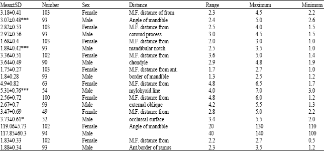 | |
| *: p<0.05, **: p<0.01, ***: p<0.001, MF: Mandibular foramen | |
| Table 2: | Statistical features of females and males of present study |
DISCUSSION
Results of present study revealed that location of MF relative to other bony landmarks had variations. These results are similar to Afsar et al. (1998) and Nicholson (1985) investigations.
MF determination is very important for anesthesia of IA nerve with Halseted approach.
Afsar et al. (1998) and Nicholson (1985) investigations were performed on Mandibular bones samples from adult persons whom nationality were from East Indian and some groups races of Canadian residences country respectively. Population of Afsar et al. (1998) studies were Black and Hispanic and Caucasian and Asian thus there were high variation in both studies.
On the other hand in Afsar et al. (1998) study, age of the population was between 10.2 and 37.5 years but, in present investigation the age was above eighteen years old. we postulated that growth of mandible interfere with results of present investigation, so we omitted the cases below 18 years old, but high variation of results were observed. It is concluded that:
| • | There were variation of MF in persons of a race and also these variations didn’t concern to the race |
| • | MF variations did not concern to mandibular growth |
We have been used panorex X-Ray film method instead of dissection method for determination of MF location, so the results could applicable for clinical cases (Afsar et al., 1998).
Mandibular foramen location can’t determine regarding other landmarks since present investigation have been revealed a high range of results. It seems present investigation is the first study on comparison of Mandibular bone between females and males. Only available data of mandible regarding sexual difference is internal inclination of the angle in female and external inclination in male (Soames, 1995). Sexual dimorphism of human skeletal mainly observed in pelvic girdle bones. There are also exist in skull to lesser extent (Soames, 1995).
Dimorphism in other skeletal bone reported (Riepert et al., 1996). In present study panorex X-Ray approach were used and measurements were exact, thus it could reveal difference of Mandible between males and females. Some respects consist of genetically differences and hormonal factors and race differences are the causes of these difference in present investigation. Results of present study revealed mandibular foramen variations related to other landmarks and dimorphism between mandibular bones of persons above eighteen years we suggest further investigations on under eighteen years persons.
ACKNOWLEDGMENTS
We wish to thank the Vice chancellor of Research in Medical Faculty of Shaheed Beheshti Medical University for financial support and Dr. Tavakoli and Mrs. Hosseini for providing X-rays films.
REFERENCES
- Afsar, A., D.A. Haas, P.E. Rossovw and R. Wood, 1998. Radiographic localization of mandibular anesthesia landmarks. Oral Surgery Oral Med. Oral Pathol. Oral Radiol. Endod., 66: 234-241.
PubMed - Kaffe, I., L. Ardekian, I. Gelerenter and S. Taicher, 1994. Location of mandibular foramen in panoramic radiographs. Oral. Surg. Oral. Med. Oral. Pathol., 78: 662-669.
PubMed - Kaafman, E., P. Weinstein and P. Milgrom, 1984. Difficulties in achiving local anesthesia. J. Am. Dent. Assoc., 108: 205-208.
PubMed - Nicholson, M.L., 1985. A study of the position of the mandibular foramen in the adult human mandible. Anat. Rec., 212: 110-112.
PubMed - Riepert, T., T. Drechsler, H. Schile, B. Nafe and R. Mattern, 1996. Estimation of sex on the basis of radiographs of calcaneus. Forensic Sci. Int., 77: 133-140.
PubMed